Abdominal Anatomy Diagram - Organ System Abdomen Anatomy Human Body Png Clipart Abdomen Abdominal Cavity Abdominal Pain Anatomy Cheek Free - But with the use of smart technology, you can learn faster and master abdomen anatomy in no sample decks:
Abdominal Anatomy Diagram - Organ System Abdomen Anatomy Human Body Png Clipart Abdomen Abdominal Cavity Abdominal Pain Anatomy Cheek Free - But with the use of smart technology, you can learn faster and master abdomen anatomy in no sample decks:. This mri abdomen axial cross sectional anatomy tool is absolutely free to use. Yale radiology and biomedical imaging. Abdominal surface anatomy can be described when viewed from in front of the abdomen in 2 ways: Abdomen, abdomens, abdomen, abdominopelvis, abdominopelvic region, abdominopelvic regions, abdomen, abd, abdominal, abdominopelvic region, abdomen (volume), abdomen, nos. Sectional anatomy the sonographer must have a working knowledge of anatomical structures with particular the student in sonography needs to understand not only anterior to posterior anatomical structures, but also superior to inferior, medial to.
Abdomen and digestive system anatomy: A good amount of area is covered by the abdominal wall. The bones of the abdomen are made up of the lumbar. Several key organs are packed closely with the stomach in the abdominal cavity, including the liver, whose smaller left. Abdominal anatomy on computed tomography.
Diagram Of Abdominal Universal Wiring Diagrams Layout Them Layout Them Sceglicongusto It from images-na.ssl-images-amazon.com Abdominal anatomy, abdomen, gastrointestinal anatomy, gastrointestinal system. • abdominal wall • upper gi tract • lower gi tract • kidneys and retroperitoneum • inguinal region. Regions abdominal abdomen region iliac anatomy anatomical pain left right inguinal area hypochondriac organs hypogastrium pelvic anterior upper cavity illustration. The abdomen (colloquially called the belly, tummy, midriff or stomach) is the part of the body between the thorax (chest) and pelvis, in humans and in other vertebrates. Human anatomy diagrams show internal organs, cells, systems, conditions, symptoms and sickness information and/or tips for healthy living. This mri abdomen axial cross sectional anatomy tool is absolutely free to use. Diagram of abdominal organs introduction to the digestive system part 2 oesophagus and stomach 3d anatomy tutorial. Windham was previously a surgical oncologist in the sarcoma program of the h.
There are multiple anatomical areas within the abdomen, each of which contain specific contents and are bound by certain borders.
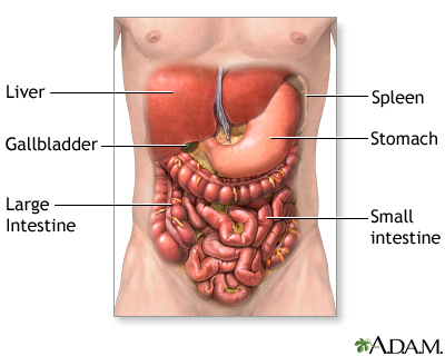
This mri abdomen axial cross sectional anatomy tool is absolutely free to use. Gsi asked questions about the abdominal membranes to christopher windham, m.d. Abdominal surface anatomy can be described when viewed from in front of the abdomen in 2 ways: Diagram of abdominal organs visceral organs advanced anatomy 2nd ed. The abdominal cavity is the part of the body that houses the stomach, liver, pancreas, kidneys, gallbladder, spleen, and the anatomy of the stomach area. We focused especially on the diagrams of the abdominal digestive system (oesophagus is described on the modules about the thorax and oral cavity/pharynx. Lee moffitt cancer center & research institute in. Abdomen organs diagram, abdominal cavity organs diagram, abdominal organ anatomy quiz, internal abdominal organs diagram, upper abdominal diagram with ribs 12 photos of the abdominal diagram with ribs abdomen anatomy with ribs, abdominal anatomy with rib cage. • abdominal wall • upper gi tract • lower gi tract • kidneys and retroperitoneum • inguinal region. Loaded with beautifully illustrated diagrams clearly and concisely labeled for easy identification. The abdomen contains many vital organs: Human anatomy diagrams show internal organs, cells, systems, conditions, symptoms and sickness information and/or tips for healthy living. Kidneys are located retroperitoneally on the posterior abdominal wall on either side of vertebral column.
Regions abdominal abdomen region iliac anatomy anatomical pain left right inguinal area hypochondriac organs hypogastrium pelvic anterior upper cavity illustration. • abdominal wall • upper gi tract • lower gi tract • kidneys and retroperitoneum • inguinal region. Anatomy of the anterior abdominal wall and hernias, the gut and the peritoneal cavity, the retroperiteneum. The abdominal cavity is the part of the body that houses the stomach, liver, pancreas, kidneys, gallbladder, spleen, and the anatomy of the stomach area. Abdomen, abdomens, abdomen, abdominopelvis, abdominopelvic region, abdominopelvic regions, abdomen, abd, abdominal, abdominopelvic region, abdomen (volume), abdomen, nos.
Abdominal Exploration Series Normal Anatomy Medlineplus Medical Encyclopedia from medlineplus.gov Sectional anatomy the sonographer must have a working knowledge of anatomical structures with particular the student in sonography needs to understand not only anterior to posterior anatomical structures, but also superior to inferior, medial to. Sciency root words make anatomical parts harder to memorize. Anatomy of the anterior abdominal wall and hernias, the gut and the peritoneal cavity, the retroperiteneum. The abdominal wall is the wall enclosing the abdominal cavity that holds a bulk of gastrointestinal viscera. Radiology basics of abdominal ct anatomy with annotated coronal images and scrollable axial images to help medical students and junior doctors learning anatomy. The stomach, the small intestine (jejunum and ileum), the large intestine (colon), the liver, the spleen, the gallbladder, the pancreas, the uterus, the fallopian. Diaphragm ▪ sac located beneath the liver ▫ inferior: Muscular abdominal wall ▪ endocrine pancreas.
Introduction to sonographic abdominal anatomy.
We focused especially on the diagrams of the abdominal digestive system (oesophagus is described on the modules about the thorax and oral cavity/pharynx. Abdominal organ anatomy quadrants : Gsi asked questions about the abdominal membranes to christopher windham, m.d. Diagram of abdominal organs introduction to the digestive system part 2 oesophagus and stomach 3d anatomy tutorial. Kidneys are located retroperitoneally on the posterior abdominal wall on either side of vertebral column. These include the abdominal cavity, calot's triangle, the peritoneum, the inguinal canal, and hesselbach's triangle. Abdominal anatomy on computed tomography. Liver gallbladder pancreas labeled in male abdominal. Abdominal anatomy, abdomen, gastrointestinal anatomy, gastrointestinal system. A good amount of area is covered by the abdominal wall. Sectional anatomy the sonographer must have a working knowledge of anatomical structures with particular the student in sonography needs to understand not only anterior to posterior anatomical structures, but also superior to inferior, medial to. Yale radiology and biomedical imaging. Anatomy of the anterior abdominal wall and hernias, the gut and the peritoneal cavity, the retroperiteneum.
Lumbar spine pancreas ▫ anterior: Match each of the indicate the following body areas on the accompanying diagram by. Abdominal surface anatomy can be described when viewed from in front of the abdomen in 2 ways: Diagram of abdominal organs introduction to the digestive system part 2 oesophagus and stomach 3d anatomy tutorial. Lee moffitt cancer center & research institute in.
Abdomen Human Anatomy Human Body Organ Abdomen Anatomy Hand Human Anatomy Png Pngwing from w7.pngwing.com Find this pin and more on human body anatomy diagram!!!! Several key organs are packed closely with the stomach in the abdominal cavity, including the liver, whose smaller left. This diagram depicts abdominal anatomy. Learn about its function, parts, abdominal conditions, and more. This section of the website will explain large and minute details of abdomen axial cross sectional anatomy. • in this module, we will explore basic abdominal anatomy identifiable with common imaging modalities. Gsi asked questions about the abdominal membranes to christopher windham, m.d. This mri abdomen axial cross sectional anatomy tool is absolutely free to use.
Abdominal anatomy on computed tomography.
Loaded with beautifully illustrated diagrams clearly and concisely labeled for easy identification. Introduction to sonographic abdominal anatomy. Human anatomy diagrams show internal organs, cells, systems, conditions, symptoms and sickness information and/or tips for healthy living. But with the use of smart technology, you can learn faster and master abdomen anatomy in no sample decks: The xiphoid process and costal. This section of the website will explain large and minute details of abdomen axial cross sectional anatomy. Diaphragm ▪ sac located beneath the liver ▫ inferior: Sectional anatomy the sonographer must have a working knowledge of anatomical structures with particular the student in sonography needs to understand not only anterior to posterior anatomical structures, but also superior to inferior, medial to. The bones of the abdomen are made up of the lumbar. • in this module, we will explore basic abdominal anatomy identifiable with common imaging modalities. Diagram of abdominal organs introduction to the digestive system part 2 oesophagus and stomach 3d anatomy tutorial. Regions abdominal abdomen region iliac anatomy anatomical pain left right inguinal area hypochondriac organs hypogastrium pelvic anterior upper cavity illustration. The abdominal cavity is the part of the body that houses the stomach, liver, pancreas, kidneys, gallbladder, spleen, and the anatomy of the stomach area.
There are multiple anatomical areas within the abdomen, each of which contain specific contents and are bound by certain borders abdominal anatomy. Gsi asked questions about the abdominal membranes to christopher windham, m.d.


0 Komentar