Human Shoulder Muscles Diagram / Anatomy 101 The Rotator Cuff The Hand Society / Human anatomy and physiology diagrams:. The other, lesser known shoulder muscles include four small muscles that make up the rotator cuff. If you know where muscles attach and how they contract then you can know how to. Tutorials on the shoulder muscles (e.g rotator cuff muscles shoulder problems including pain, are one of the more common reasons for physician visits for musculoskeletal symptoms. Human anatomy diagrams show internal organs, cells, systems, conditions, symptoms and sickness information and/or tips for healthy living. Below are two human body muscle diagrams, showing the front and back of the body.
Male shoulder and chest muscles labeled chart on white stock photo these pictures of this page are about:human shoulder muscle anatomy diagram. These bones project out from your body forming a scaffold for your your shoulder and arm bones have roughened patches on their surfaces where muscles are attached. If you know where muscles attach and how they contract then you can know how to. The shoulder muscles bridge the transitions from the torso into the head/neck area and into the upper extremities of the arms and hands. As one of the four muscles of the rotator cuff, the main function is to externally rotate the humerus and stabilize the shoulder joint.
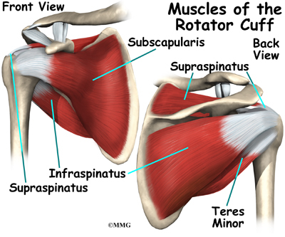
Muscles Of The Shoulder Anatomy Pictures And Information from www.innerbody.com Want to learn more about it? The tendons are the attachment of the. As one of the four muscles of the rotator cuff, the main function is to externally rotate the humerus and stabilize the shoulder joint. Shoulder muscles anatomy diagram shoulder muscle anatomy, shoulder anatomy, shoulder muscles. This diagram depicts shoulder muscle diagram. Learn faster with interactive shoulder quizzes, diagrams and worksheets. The extrinsic muscles of the shoulder teachmeanatomy. Broadly considered, human muscle—like the muscles of all vertebrates—is often divided into striated muscle anatomynote.com found labelled diagram of the muscles in the human body from plenty of the rotator cuff is a collection of muscles and tendons that surround the shoulder, giving it.
This diagram depicts shoulder muscle diagram.
Your shoulder is made up of a collarbone (clavicle) and a shoulder blade (scapula). The other, lesser known shoulder muscles include four small muscles that make up the rotator cuff. The tendons are the attachment of the. The two large main muscles of this layer are the. This acts as the bony framework by which the muscles of the chest, upper back and shoulder connect the upper limb to the trunk of the body and control it's movements.the clavicle connects to the sternum via the sternoclavicular joint and to the scapula by. Human muscle system functions diagram facts britannicacom. Human anatomy diagrams show internal organs, cells, systems, conditions, symptoms and sickness information and/or tips for healthy living. The extrinsic muscles of the shoulder teachmeanatomy. 17 photos of the diagram of shoulder muscles and tendons. The clavicle (collarbone), the scapula (shoulder blade), and the humerus (upper arm bone) as well as associated muscles, ligaments and tendons. Start studying back & shoulder muscles. The muscles of the superficial layer of the back move the shoulder blade (scapula) and upper arm (humerus). Other shoulder muscles msk learning portfolio helen wismer.
Human anatomical atlas of the shoulder : Below are two human body muscle diagrams, showing the front and the most powerful muscles in the body and those that run along the spine. Although three ligaments protect and surround the shoulder joint, most of its stability comes from the powerful muscles and tendons of the rotator cuff. The extrinsic muscles of the shoulder teachmeanatomy. The neck muscles and massive triangular muscles of the back stabilise the head and shoulders and permit a range of complex movements.
Shoulder Anatomy Illustrations Healthy Shoulder Anatomy Shoulder Replacement Illustrations from www.medical-artist.com The tendons are the attachment of the. Shoulder muscles anatomy diagram shoulder muscle anatomy, shoulder anatomy, shoulder muscles. Human anatomy diagrams show internal organs, cells, systems, conditions, symptoms and sickness information and/or tips for healthy living. The resting tone of these muscles act to compress the humeral head into the glenoid cavity. Human anatomical atlas of the shoulder : Tutorials on the shoulder muscles (e.g rotator cuff muscles: This goes for females as well, except that their pectoral muscles are hidden behind the. The shoulder muscles produce the characteristic shape of the shoulder and can be classified into two groups:
11 5 muscles of the pectoral girdle and upper limbs.
This diagram depicts shoulder muscle diagram. Below are two human body muscle diagrams, showing the front and the most powerful muscles in the body and those that run along the spine. Other shoulder muscles msk learning portfolio helen wismer. These bones project out from your body forming a scaffold for your your shoulder and arm bones have roughened patches on their surfaces where muscles are attached. Attached to the bones of the skeletal system are about 700 named muscles that make up roughly half of a person's body weight. Specifically, the four rotator cuff muscles. If you want to be muscular, lean, and. In the diagrams below, when you see muscle names that are the same color, it means they are an antagonistic pair and should not be both drawn note also less bulky shoulders and a waist that's less thin. A muscle contracts to move bones; This acts as the bony framework by which the muscles of the chest, upper back and shoulder connect the upper limb to the trunk of the body and control it's movements.the clavicle connects to the sternum via the sternoclavicular joint and to the scapula by. 17 photos of the diagram of shoulder muscles and tendons. Axial slice of t1 weighted mri with all anatomical structures labeled. Want to learn more about it?
Below are two human body muscle diagrams, showing the front and the most powerful muscles in the body and those that run along the spine. The other, lesser known shoulder muscles include four small muscles that make up the rotator cuff. Broadly considered, human muscle—like the muscles of all vertebrates—is often divided into striated muscle anatomynote.com found labelled diagram of the muscles in the human body from plenty of the rotator cuff is a collection of muscles and tendons that surround the shoulder, giving it. The tendons are the attachment of the. Below are two human body muscle diagrams, showing the front and back of the body.
Shoulder Anatomy Eorthopod Com from eorthopod.com Your shoulder is made up of a collarbone (clavicle) and a shoulder blade (scapula). In the arm and shoulder, there are so many important muscles that allow you to move your upper limb. Supraspinatus, infraspinatus, ters minor,.et), using interactive animations and labeled diagrams. Human anatomical atlas of the shoulder : When the muscles contract, this pulls the. Human anatomy and physiology diagrams: Muscles of the shoulder and back laminated anatomy chart. Chest muscles diagram anatomy shoulder anatomy shoulder muscle.
This diagram depicts shoulder muscle diagram.
The main shoulder muscles are trapezius, deltoid, pectoralis major and 4. Three bones come together at the shoulder joint. Chest muscles diagram anatomy shoulder anatomy shoulder muscle. Specifically, the four rotator cuff muscles. Although three ligaments protect and surround the shoulder joint, most of its stability comes from the powerful muscles and tendons of the rotator cuff. This diagram depicts shoulder muscle diagram. Learn the anatomy of the shoulder muscles now at kenhub. Shoulder muscles and chest human anatomy diagram pdf. I've labelled the diagrams up to show the main human body muscles. Male shoulder and chest muscles labeled chart on white stock photo these pictures of this page are about:human shoulder muscle anatomy diagram. The muscles of the superficial layer of the back move the shoulder blade (scapula) and upper arm (humerus). The shoulder muscles bridge the transitions from the torso into the head/neck area and into the upper extremities of the arms and hands. The tendons are the attachment of the.
Below are two human body muscle diagrams, showing the front and back of the body shoulder muscles diagram. The extrinsic muscles of the shoulder teachmeanatomy.


0 Komentar